Arizona's Vision Eye Care Center

Several eye conditions produce symptoms nearly identical to macular degeneration, including blurred central vision, distorted straight lines, and dark spots in the visual field. Without a thorough diagnostic evaluation, these conditions are frequently misidentified, which delays the correct treatment and puts long-term vision at risk. Understanding which conditions overlap with macular degeneration helps patients in Phoenix, AZ recognize why an accurate diagnosis matters and why symptoms alone are never enough to determine the cause.
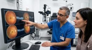
Which Conditions Are Most Commonly Confused With Macular Degeneration?
Central serous chorioretinopathy, diabetic macular edema, macular holes, epiretinal membranes, and inherited macular dystrophies such as Stargardt disease are the conditions most frequently mistaken for macular degeneration. Each one affects the macula or central retina, produces overlapping visual symptoms, and requires specific diagnostic imaging to distinguish from age-related macular degeneration (AMD).
Central Serous Chorioretinopathy
Central serous chorioretinopathy (CSC) causes fluid to accumulate beneath the retina, distorting central vision in a way that closely resembles early AMD. It most commonly affects men between the ages of 30 and 50, which is a key distinguishing factor since AMD typically presents in patients over 55. Stress and corticosteroid use are known triggers. Because the visual distortion and central blurring are nearly identical to AMD symptoms, CSC is one of the most common misdiagnoses without imaging confirmation. Optical coherence tomography (OCT) clearly differentiates the two by revealing the subretinal fluid pattern specific to CSC.
Diabetic Macular Edema
Diabetic macular edema (DME) occurs when diabetes-related vascular damage causes fluid to leak into the macula, swelling the central retinal tissue and reducing visual acuity. Patients with DME often report blurred or wavy central vision, which mirrors AMD presentation. The critical difference is systemic context: DME develops in patients with diabetic retinopathy, while AMD is driven by age-related degeneration of the retinal pigment epithelium. A patient without a known diabetes diagnosis may not immediately connect their vision changes to blood sugar, making the distinction harder to recognize without a full medical and ocular history.
Other Eye Diseases That Mimic Macular Degeneration Symptoms
Several additional retinal conditions produce central vision loss that overlaps with AMD, particularly in patients who have not yet received a formal diagnosis.
Macular Hole and Epiretinal Membrane
A macular hole is a small break in the macula that causes a distinct central blind spot, often described as a dark or missing area in the center of vision. An epiretinal membrane, sometimes called a macular pucker, is a thin layer of scar tissue that forms over the macula and causes central distortion and blurring. Both conditions are structural rather than degenerative, meaning they result from physical changes to the retinal surface rather than the progressive cell loss seen in AMD. OCT imaging distinguishes these structural abnormalities from AMD with high precision.
Stargardt Disease and Inherited Macular Dystrophies
Stargardt disease is the most common inherited macular dystrophy, typically presenting in children and young adults with progressive central vision loss. Because the central vision decline and macular changes visible on examination resemble AMD, Stargardt disease is sometimes misidentified in younger patients who present without a family history. Other inherited dystrophies, including Best disease and pattern dystrophy, produce similar macular changes. Genetic testing and specialized retinal imaging are required to confirm an inherited cause and rule out AMD.
Recognizing that these conditions exist alongside AMD is only part of the picture. A comprehensive dilated eye exam is what separates a symptom pattern from an accurate diagnosis, because the clinical tools used to evaluate the retina reveal structural and vascular details that symptoms alone cannot.
Why These Misdiagnoses Happen and What They Mean for Your Care
Misdiagnosis occurs because the macula is a small, highly specialized region of the retina, and damage to it produces a limited set of visual symptoms regardless of the underlying cause. Central blurring, metamorphopsia (distorted straight lines), and central scotomas appear across multiple conditions. Without imaging, the symptom profile of age-related macular degeneration looks nearly identical to CSC, DME, or a macular hole on patient report alone.
How a Comprehensive Eye Exam Differentiates These Conditions
A dilated fundus examination, OCT imaging, fundus photography, and fluorescein angiography each reveal different layers of retinal structure and vascular behavior. OCT shows retinal thickness, fluid pockets, and structural integrity. Fluorescein angiography maps blood vessel leakage patterns. Together, these tools allow an eye care provider to identify the specific pathology affecting the macula. In cases where the findings suggest a retinal condition beyond the scope of primary eye care, when a referral to a retinal specialist is appropriate becomes the next clinical decision, ensuring the patient receives the right level of care without delay.
Conclusion
Multiple retinal conditions share the visual symptoms of macular degeneration, and distinguishing between them requires diagnostic imaging, not symptom assessment alone. The correct diagnosis determines the correct treatment, and the two are not interchangeable.
For patients in Phoenix, AZ experiencing central vision changes, early evaluation is the single most important step toward protecting long-term visual function and avoiding the consequences of a missed or delayed diagnosis.
At Arizona's Vision Eye Care Center, our team guides patients through every step of the diagnostic process with clarity, precision, and compassionate care.
Frequently Asked Questions
Can central serous chorioretinopathy be mistaken for macular degeneration?
Yes. Both conditions cause central vision distortion and blurring. CSC primarily affects younger men and involves subretinal fluid, while AMD involves retinal pigment epithelium degeneration. OCT imaging reliably distinguishes the two.
What is the difference between macular degeneration and diabetic macular edema?
AMD results from age-related retinal cell degeneration. Diabetic macular edema is caused by fluid leakage from diabetes-damaged blood vessels. Both affect central vision, but their causes, risk factors, and treatments differ significantly.
How do doctors tell macular degeneration apart from other retinal diseases?
Optical coherence tomography, fluorescein angiography, and dilated fundus examination reveal the specific structural and vascular changes associated with each condition, allowing precise differentiation that symptoms alone cannot provide.
Can young people be diagnosed with macular degeneration?
Age-related macular degeneration is rare in patients under 55. Central vision loss in younger patients is more likely caused by inherited dystrophies such as Stargardt disease, CSC, or other retinal conditions requiring separate evaluation.
What symptoms should prompt an immediate eye exam?
Sudden blurred central vision, distorted straight lines, a dark or missing spot in the center of your visual field, or rapid changes in color perception all warrant prompt evaluation by an eye care professional.

Dr. Page is a licensed Doctor of Optometry, and a long time resident of Ahwatukee Arizona. He graduated from the New England College of Optometry in Boston in 1990. He moved to Phoenix to serve the community in both his clinic and by becoming a community activist.
He is deeply involved in supporting the local athletic teams, the arts, pet adoption and children. He is a charter member of the Chamber of Commerce.
After watching his father lose vision in his right eye he dedicated his career to finding a way to stopping eyes from getting worse. He is the founder of the Invisalens eye retainer and a best selling author for his book: Freedom! The Smart Parents Guide to Helping Your Child See a Better Life.
He specializes in helping his patients see better with eyeglasses, contact lenses, nutrition and genetics.
Words can't describe what our eyes bring to life!